Ads 468x60px

Featured Posts Coolbthemes

Trypanosomiasis

The trypanosomiases are a disease complex that affects man and his livestock in Africa, Asia and South America. ILRAD is concerned with those trypanosomiases transmitted by tsetse flies, which threaten the lives of some 30% of an estimated population of 150 million cattle in 37 countries of Africa, as well as comparable numbers of small ruminants. Annual losses in meat production alone are estimated at US$5 billion. This economic deprivation is exacerbated by losses in milk production, tractive power, waste products that provide natural fuel and fertilizer and secondary products such as clothing and hides. In addition, 50 million people are currently exposed to the risk of contracting human trypanosomiasis, known as sleeping sickness.

Trypanosomiasis is caused by blood-dwelling protozoan parasites called trypanosomes, which infect man, cattle, sheep, goats, pigs, horses and camels. Wild animals can also be infected and serve as reservoirs of infection. Trypanosomes are transmitted by tsetse flies (Glossinidae) and other biting insects, which ingest the parasites in blood meals taken from infected animal hosts (Figure 12). In the tsetse fly the parasites multiply in the midgut or in the proboscis and undergo a developmental cycle culminating in the generation of metacyclic trypanosomes, which are infective to the mammalian host. Metacyclic forms are introduced into a new host through the saliva of the tsetse fly during feeding. In the mammalian host the parasites develop in the tissues and blood, causing anaemia, reduced productivity, reproductive disorders and death.
Figure 12. Schematic diagram showing the life cycles of the three major tsetse-transmitted trypanosome species in Africa.

Three major species of trypanosomes of veterinary importance are studied at ILRAD. These are Trypanosoma congolense, Trypanosoma vivax and Trypanosoma brucei brucei. Biological aspects of Trypanosoma evansi, which belongs to the same Trypanozoon group as T. brucei but which is transmitted by biting flies other than tsetse, are also studied at ILRAD. Trypanosoma simiae, which is closely related to T congolense, is primarily a parasite of pigs but is frequently found in trypanosome isolates taken from ruminants and tsetse flies in the field. Two further subspecies of T. brucei, T. brucei rhodesiense and T. brucei gambiense infect man and are reservoirs of infection in various types of wild animals.

In addition to there being many species of trypanosomes, a phenomenon called antigenic variation exhibited by each species helps to induce chronic infections in the hosts the parasites infect. Trypanosomes are covered by a dense coat made up of glycoprotein molecules, called variable surface glycoproteins (VSGs) (Figure 13). A trypanosome population causing infection in a single susceptible host animal can vary these surface antigens in a matter of a few days. Although the host usually generates a good antibody response to trypanosomes expressing a particular variable antigenic type (VAT), the large number of VATs that can be expressed within a single infection, estimated to be 300-1000 for T. brucei, causes chronic infections. Long-term infections in turn help ensure the transmission of the parasites to other animals and increase the severity of the pathogenesis, particularly anaemia, caused by trypanosome infection.
Figure 13. Schematic diagram of a trypanosome of the Trypanosoma brucei group in its intermediate bloodstream form, illustrating the major organelles.

It was initially believed that study of the antigenic variation displayed by trypanosomes would best elucidate how trypanosomes maintain themselves in the host animal. This information could then be used to develop vaccines to control or prevent the infection. But detailed studies of antigenic variation carried out at ILRAD and elsewhere over the last several years suggest that the number of VSGs expressed by trypanosomes is too great to make a vaccine based on the administration of a single, or even a few, VATs broadly effective. The total number of antigens expressed by any stock of trypanosomes, called a repertoire, is very large. These antigenic repertoires vary not only among but also within species: isolates in one area of Africa often differ in their antigenic repertoire from isolates in other areas. These distinct antigenic repertoires of a single species are known as serodemes. The existence of serodemes considerably enlarges the number of VATs encountered in the field.

Traditional management of trypanosomiasis has relied on chemotherapeutic drugs to treat infections in livestock and on insecticides to control the tsetse vector. Because the widespread use of insecticides is environmentally damaging, attempts have recently been made to control the disease by combining the strategic use of insecticide-impregnated traps and screens, to reduce the tsetse populations, with chemotherapeutic treatment for the animals that succumb to the lowered challenge. This approach, when followed by close monitoring of the tsetse population and the disease status of livestock, has successfully controlled trypanosomiasis in a few trial areas, but these methods are too difficult to apply in most parts of Africa. Furthermore, relying on one or two drugs to treat infected livestock increases the possibility that the parasites will develop resistance to the drugs.

ILRAD is therefore taking an alternative approach and is seeking primarily immunological solutions to the problems posed by trypanosomiasis. The complexity of the antigenic variation exhibited by the parasite has necessitated that the research goals of the trypanosomiasis program be divided into short and long term. In the short term ILRAD is studying ways to develop more accurate tests to diagnose trypanosome infection in livestock, new ways with which to use the currently available trypanocides that will reduce the possibility of inducing parasite resistance to the drugs, and the reasons why some breeds of cattle are able to withstand trypanosome infection better than others. In the long term ILRAD is conducting research in two main areas. It is studying the responses ruminant hosts make to trypanosome infection, with the aim of enhancing normal mechanisms of resistance in livestock, and it is scrutinizing molecules and processes of the parasite in a search for key elements or activities that can be attacked with drugs without adversely affecting the host.

Short-term strategies for trypanosomiasis control

Tests to diagnose infections

To detect and identify trypanosome infections in tsetse flies, domestic livestock and man, ILRAD scientists for several years have been developing diagnostic tests that use monoclonal antibodies in enzyme-linked immunosorbent assays (ELISAs). Reagents are also available to identify trypanosomes of the subgenera Trypanozoon, Nannomonas and Duttonella. In 1988 new monoclonal antibodies were developed to identify protein rather than carbohydrate epitopes of T. vivax and T. congolense membrane proteins. These antibodies will reduce the likelihood of the ELISA giving false positive results. Furthermore, by detecting target proteins, the antibodies will enable scientists to identify fragments of DNA encoding the trypanosomal antigens in libraries of complementary DNA-DNA copied from messenger RNA of the various trypanosome species. Recombinant antigens could then be synthesized in great quantity for use in inhibition ELISAs, which may be more sensitive in detecting infections than the technique presently used. In 1988 the trypanosomiasis program began to investigate the synthetic production of the diagnostic antigen for T. brucei.

The assays in their present form are now being tested for accuracy and sensitivity in national laboratories in ten African countries in collaboration with the World Health Organization and a joint division of the Food and Agricultural Organization of the United Nations and the International Atomic Energy Agency. Work conducted in collaboration with the University of Brussels and national laboratories in Africa and Asia has shown that the test to diagnose T. brucei can also be used to diagnose T. evansi infections in domestic livestock. Trypanosoma evansi is an economically important pathogen in Africa, Asia and Latin America. The efficacy of this assay in detecting T. evansi infections in buffalo, pigs and horses will be tested in Indonesia in a collaborative program run by ILRAD and research institutes in Indonesia and Australia.

Molecular probes to identify species

Our understanding of the epidemiology of both animal and human trypanosomiasis is improved by an ability to identify more precisely the trypanosome species carried by tsetse flies. To this end ILRAD has produced DNA probes to detect differences among parasite species and subspecies at the genomic level. These highly sensitive probes can be used in the laboratory to distinguish repetitive DNA sequences that recognize specific species and subspecies of pathogenic trypanosomes. The probes also distinguish differences among trypanosome populations of the same species collected from different areas. When the probes were tested on field materials collected in Kenya in collaboration with the International Centre of Insect Physiology and Ecology (Nairobi), it was discovered that although the probes are species-specific and detect low numbers of trypanosomes in infected tsetse flies, the probes do not detect all isolates of a given species. These results demonstrate that genetic differences occur within species; attempts will now be made to identify DNA sequences common to all populations of a given trypanosome species.

All the genetic probes in use today are radioactively labelled. Because most laboratories in Africa lack radioisotope facilities, these probes are not widely employed. Their use would be greatly promoted if the radioisotope were replaced with simple chemical groups that could be detected immunologically. ILRAD scientists have addressed this problem by producing derivatives of the probes with dinitrophenol so that positive hybridization using the repetitive sequence DNA probes can be revealed by an enzyme-labelled anti-dinitrophenol antibody. Although the reproducibility of the derivatization depends critically on the purity of dinitrobenzaldehyde, the initial labelling agent, dinitrophenol-labelled probes can detect as little as 1-5 pg of purified DNA, which demonstrates a sensitivity similar to that of the radiolabelled probes and probably sufficient for detecting trypanosomes in tsetse.

Future work at ILRAD in this area will attempt to validate the diagnostic assays and to increase their sensitivity where that is desirable. ILRAD staff will also work with scientific organizations to develop forms of the tests that can be widely and easily used in the areas of the world affected by trypanosomiasis.

Chemotherapy

One of the principal objectives of the work conducted at ILRAD on chemotherapy and chemoprophylaxis is to develop assays to quantify the levels of drugs in the blood and tissue fluids of treated livestock. Two kinds of such assays chemical and biological have been produced. The former, which requires sophisticated chemical techniques, will be used to quantify biological assays based on trypanosome culture systems. The simpler biological assays should prove useful to national laboratories.

In vitro assays of drug resistance

ILRAD scientists have shown that incorporating drugs in culture medium can inhibit the growth and development of trypanosomes in vitro, depending upon the sensitivity of the trypanosome population to the drugs used. Cultures of the bloodstream forms of the parasite were used in the first experiments in this area. Clear differences in Berenil-induced growth inhibition were observed (Figure 14). Bloodstream forms of fresh field isolates, however, are difficult to adapt to cultures. Cultures of procyclic forms of the trypanosome the noninfective, multiplying forms of the parasite equivalent to the forms found in the tsetse midget of T. brucei and T. congolense seem to be established more reliably, and tube assays have been developed for isolating and testing T. congolense and T. brucei for drug sensitivity.

Figure 14. Graph showing the comparative growth inhibition of drug-susceptible (IL 1.4) and drug-resistant (CP 547/r and CP 2469) trypanosomes in the presence of diminazene aceturate (Berenil). Trypanosomes were incubated at 37°C in 4% C02 in air for 24 hr in culture medium containing various drug concentrations. Growth was determined with a Coulter Counter and compared with control cultures.
Blood samples containing trypanosomes are mixed with a culture medium and incubated overnight at 27°C in a plastic centrifuge tube. The trypanosomes in the supernatant are removed and transferred to culture flasks to allow the procyclic forms to develop and grow further. All trypanosome stocks were successfully isolated by this procedure in a medium based on the composition of tsetse haemolymph to which supplements had been added.
The growth of procyclic forms of drug-sensitive trypanosome stocks was inhibited when these were propagated for ten days in the presence of 1 ng/ ml of isometamidium chloride. In cultures of sensitive trypanosomes, the drug induced an increase in cell size, degradation of the kinetoplast and multinucleate forms to develop. Differences in growth inhibition of procyclic forms between resistant and sensitive stocks also occurred after incubation for 48 hours with 1�100 ng/ ml isometamidium chloride. Only minor differences, however, were observed when procyclic forms of resistant and sensitive T. b. brucei were cultivated with diminazene aceturate. Thus, whereas inhibition of the growth of procyclic forms can be used to test trypanosome stocks for their levels of resistance to isometamidium chloride, the same assay appears to be inappropriate for diminazene aceturate.
An alternative method of testing trypanosome populations for resistance to both isometamidium chloride and diminazene aceturate is the drug incubation infectivity test. Cultures of trypanosomes are incubated at 37° C in 4% carbon dioxide in air for 24 hours in the presence of a drug or plasma from drug-treated animals. Control cultures incubated with 1 % distilled water instead of a drug are incubated under the same conditions. After incubation, an aliquot from each culture is inoculated into mice. The mice are then screened for 30 days for trypanosome infections.
None of the drug-sensitive trypanosome stocks were able to infect the mice after incubation with 1 ng/ ml isometamidium chloride, but all resistant stocks were able to do so. Two of the isometamidium-resistant trypanosome stocks retained their infectivity after incubation with 10 or 50 ng/ ml of the drug. But when used at higher concentrations on resistant trypanosome populations, the drug increased the period between infection and the appearance of parasites in the blood of the mice.
Higher concentrations of diminazene aceturate were necessary to inhibit the infectivity of T. b. brucei. Differences in infectivity occurred when trypanosomes were incubated in the presence of 0.05�1.00 µg/ ml of diminazene aceturate. The drug-resistant trypanosome stocks retained infectivity after incubation with 1 µg/ ml of the drug. Thus, by using the drug incubation infectivity test it is possible to distinguish isometamidium-and diminazene-resistant trypanosome populations from those that are sensitive to these drugs. The test has been validated for T. brucei, T. evansi and a rodent-infective stock of T. vivax.
The stability of the drug-resistant trait has been further examined in vitro: a T. brucei stock resistant to diminazene, isometamidium, quinapyramine and mel B was grown in vitro and its sensitivity to these drugs was compared to that of a drug-sensitive trypanosome stock. There was little change in sensitivity after propagation for up to 275 days of bloodstream forms in vitro or after transformation of bloodstream forms into procyclic, epimastigote and, finally, metacyclic forms. These results suggest that drug resistance is a relatively stable characteristic even when trypanosomes are maintained in vitro in the absence of drugs.
Significant progress was made in cultivating certain stocks of the Trypanozoon group without feeder-layer cells. This has enabled researchers to conduct several new studies, including an evaluation of trypanocides in the absence of feeder-layer cells.

Drug resistance

The development of resistance in trypanosomes to the trypanocidal drugs currently available threatens our ability to continue to control trypanosomiasis. In collaboration with the departments of Veterinary Parasitology and Physiology at the University of Glasgow, ILRAD is conducting studies to determine new ways of treating livestock that are infected with trypanosomes known to be resistant to recommended doses of trypanocidal drugs. Reports from the field have suggested that intravenous administration of isometamidium chloride has a curative effect on cattle infected with trypanosome populations that are resistant to the drug when it is administered intramuscularly. In experiments at ILRAD, however, the intravenous administration of high levels of isometamidium chloride failed to cure cattle infected with cloned populations of T. congolense that were resistant to this drug when it was administered intramuscularly.
Using highly sensitive radiochemical and chemical techniques, a relay toxicity study, carried out in 1988 by the Department of Veterinary Pharmacology at the University of Glasgow in collaboration with ILRAD and the manufacturers of isometamidium chloride, Rhone-Poulenc (UK), showed that isometamidium chloride cannot be absorbed into blood and body fluids from the gastrointestinal tract of animals that were either dosed with the drug by mouth or fed with tissues of an animal given the drug intramuscularly one week previously. This makes it unlikely that human consumption of meat from animals treated with isometamidium chloride is a significant health hazard.

Tsetse biology

Male Glossina flies sexually sterilized by gamma-irradiation carry trypanosomiasis as efficiently as fertile males. However, when tsetse are given a blood meal containing 8�12g/ml of isometamidium chloride before a blood meal containing trypanosomes, infection of the flies by mature T. vivax, T. congolense and T. b. brucei was completely suppressed. It is therefore recommended that the flies used in the sterile insect release method of tsetse control be fed on blood containing isometamidium chloride.
Various other factors were investigated to determine their influence on the maturation of the three major species of trypanosomes and their transmission by tsetse. These parameters included the species of the wild mammalian hosts and tsetse vectors of the parasites and the occurrence of pre-existing trypanosome infections in the hosts and rickettsia-like organisms in the vectors (Figure 15). In ILRAD experiments, G. m. centralis appeared to be largely unaffected by these parameters and proved the most efficient vector of trypanosomiasis.

Figure 15. Electron micrograph of part of a midgut epithelial cell from a 30-day-old non-teneral Glossina morsitans centralis, showing the presence of large numbers of rickettsia-like organisms (R) throughout the cytoplasm. Magnification = ×5,300.
Further entomological research at ILRAD shows that the developmental cycle of T. vivax in tsetse, thought to be confined to the proboscis of the fly, may extend to the cibarial-oesophageal region, the most anterior part of the digestive tract of a tsetse. Tsetse proboscides were excised at intervals beginning one hour after an infected feed and transferred to culture dishes. Parasite multiplication and full cyclical development were observed only in proboscides excised four or more hours after the infected blood meal. It thus appears that in some tsetse, development of T. vivax through its vector stages is initiated in the cibarial/ oesophageal region, from where the parasites migrate to the food canal of the proboscis, where maturation to the metacyclic forms completes the life cycle of the parasite.
Early in 1988 a tsetse control campaign conducted by the African Trypanotolerant Livestock Network was started in Côte d'Ivoire using biconical traps impregnated with cypermethrin insecticide. Initial results indicate that this control method can reduce tsetse populations by more than 98%.
A comparison of tsetse blood meals at two sites in Zaire and one site in Gabon demonstrated that Glossina tabaniformis�a species of tsetse belonging to the fusca, or forest-dwelling group�takes a considerable proportion of feeds from cattle in these areas when this species comes into contact with the cattle. Because the fusca group inhabits forest zones of West and Central Africa, most of which are distant from cattle grazing areas, it had previously been thought that G. tabaniformis contributed little to trypanosome transmission.

Trypanotolerance

Trypanotolerant livestock, principally the N'Dama breed of cattle from West Africa, offer another means of improving agricultural productivity in tsetse-infected areas. ILRAD collaborates with the International Livestock Centre for Africa (ILCA [Addis Ababa]) and national livestock ministries and development programs in West and Central Africa in the African Trypanotolerant Livestock Network. With this network and the International Trypanotolerance Centre (the Gambia), ILRAD is studying the productivity of trypanotolerant livestock under different levels of trypanosomiasis risk and is seeking economically viable ways to increase the productivity of these livestock.
ILRAD contributes to the network's activities by conducting collaborative programs on bovine genetics and trypanocide use and by helping national organizations in six countries apply standardized techniques in the collection of entomological and animal health data. A statistically significant relationship has been established at several sites between the monthly estimates of tsetse challenge and trypanosome prevalence in trypanotolerant cattle.
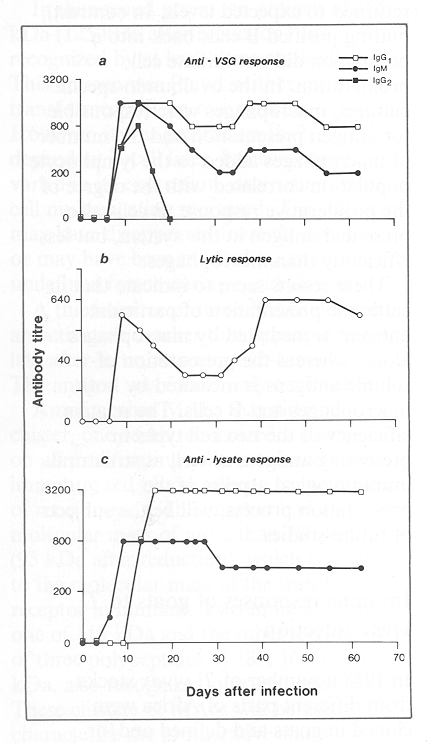
In a search for genetic markers of trypanotolerance, two polymorphic systems of bovine lymphocyte antigens were studied in 1988 in collaboration with ILCA. These systems are the major histocompatibility complex (MHC) and a more limited polymorphic system of common leucocyte antigens, which was detected in cattle only recently. The first objective of the study was to survey the MHC and common leucocyte antigen phenotypes of populations of N'Dama cattle in Zaire and the Gambia and to compare these phenotypes with corresponding profiles of trypanosensitive Boran cattle in Kenya. The second objective was to look for associations between these MHC and common leucocyte antigen phenotypes, trypanotolerance and the productivity of N'Dama cattle. Significant correlations have been found between the two classes of lymphocyte markers and the degree of resistance shown by trypanotolerant cattle exposed to trypanosomiasis by natural challenge. These provocative results, which suggest that there is a genetically selectable marker for the trypanotolerant trait, are being investigated further using larger numbers as well as family groups of cattle. The results also indicate a central role for immunity in the manifestation of the trypanotolerant trait.
ILRAD's embryo transfer experiments continued in 1988. N'Dama heifers produced at ILRAD from frozen N'Dama embryos brought in 1983 from the Gambia have since 1987 been regularly induced to superovulate using Folltropin or follicle-stimulating hormone derived from pigs. By implanting the best of the N'Dama embryos in Boran foster mothers, ILRAD has produced 24 N'Dama calves, which are used in studies of trypanotolerance and bovine genetics. ILRAD hopes to produce twin N'Dama calves by using these techniques so that research may be carried out on genetically matched animals.
The African Trypanotolerant Livestock Network will continue to evaluate the performance of trypanotolerant livestock in tsetse-infested areas of Africa, to seek ways to exploit the genetic resistance to trypanosomiasis in such livestock and to determine the most economic ways of improving the productivity of these livestock by nutritional and breeding strategies.

Long-term strategies for trypanosomiasis control

The biology of the trypanosome

The current methods used to control trypanosomiasis in cattle breeds other than N'Dama are highly susceptible to breakdown. The search for novel and more sustainable control measures for these breeds therefore forms the basis of ILRAD's long-term trypanosomiasis research program. Emphasis in this program is put on finding immunological ways to control the disease. Research is being conducted on the biology of the parasite in an attempt to discover processes unique to the parasite that could be the target of interventions. The aims of research on host-parasite interactions are to elucidate host mechanisms involved in controlling and destroying the parasite and then to enhance these mechanisms so as to reduce or alleviate the pathogenic manifestations of the disease. Research projects on basic parasite processes, such as the uptake of nutrients and the control of differentiation, were consolidated during the year and methods to cultivate trypanosomes were improved.

Antigenic variation

The variable surface glycoproteins (VSGs), which make up the surface coat of the trypanosome, bear a complex lipid-containing structure at their carboxyl end called the glycosylphosphatidylinositol, or GPI moiety. Part of this structure is responsible for anchoring the VSG molecules in the plasma membrane of the trypanosome. Unlike the protein part of VSG molecules, which differs from one VSG to another, the hydrophobic GPI anchor is similar in many of the surface coat molecules examined from T. brucei and T. congolense. Antibodies can be raised in rabbits that will react with virtually all VSGs from these trypanosome species because the antibodies bind to a cross-reacting determinant (CRD) within the hydrophobic anchor and not to the variable, protein part of the VSG molecule.
It has been suggested that the lipid portion of the anchor must be removed from membrane-form VSG (mfVSG) by an endogenous VSG-specific phospholipase C (GPI-PLC) before antibodies will bind to the CRD. Using T. congolense metacyclic forms from culture and substantiating their findings with bloodstream forms of T. congolense and T. vivax, workers at ILRAD have shown that the method of sample preparation is extremely important: the anti-CRD antibodies will bind to suitably prepared VSGs without prior removal of the lipid. However, the addition of dithiothreitol, which causes the protein part of the VSG molecule to partially unfold, was crucial in these experiments (Figure 16). It would seem, therefore, that a conformational change in the protein part of the VSG molecule is needed to expose the CRD to the antibodies before the antibodies can bind to the CRD.


Figure 16. Western blot probed with anti-CRD (cross-reacting determinant) antibodies illustrates that the binding of anti-CRD antibodies to VSG (variable surface glycoprotein) is dependent upon disulphide bond reduction: Lane A, Trypanosoma congolense metacyclics reduced with dithiothreitol; Lane B, T. congolense metacyclics without reduction; Lane C, T. congolense bloodstream forms reduced with dithiothreitol; Lane D, T congolense bloodstream forms without reduction. Metacyclic forms show a qualitative effect in anti-CRD antibody binding, bloodstream forms a quantitative effect.
Using density centrifugation of subcellular fractions of T. brucei, the enzyme responsible for removing the lipid from the hydrophobic anchor (the GPI-PLC) has been tentatively localized in a vesicle that contains both an internalized protein (bovine serum albumin) and an enzyme of the trypanosomal flagellar pocket (adenyl cyclase). The co-localization of these three reagents suggests that the GPI-PLC may reside in a vesicle near the flagellar pocket and that it may play an important role in recycling VSG.
At the molecular genetic level, metacyclic VSG genes of T. congolense IL 3000 have been cloned and expressed in the bacterium Escherichia coli. This has provided nucleotide sequence data for the VSGs of genuine metacyclic trypanosomes. The only material previously available for examination comprised bloodstream-form trypanosomes expressing cross-reactive VSGs. The genetic context in which these metacyclic VSG genes are expressed is now being examined. This is important because metacyclic trypanosomes express a constant but smaller set of VSGs than bloodstream forms belonging to the same repertoire.
Differences have already been observed between the two metacyclic VSG genes that have been cloned, mVSG1 and mVSG2. Only one copy of mVSG1 is conserved in all the developmental stages examined; at least two copies of mVSG2 exist in the metacyclic trypanosomes. Each of the metacyclic VSG genes is expressed at a different locus on chromosomes that are 2 megabases in size. One gene, mVSG1, is activated in situ; the other, mVSG2, is rearranged when it is expressed.
Work is in progress to determine the amino acid sequence and ancillary moieties of two VSGs from rodent-infective T. vivax clones. No peptide homology between the two VSGs has been demonstrated by using serological techniques and little information on the amino acid sequence has been obtained from analyses of purified VSGs. However, the importance of this work was manifested in 1988 with the discovery that, unlike the little antigenic cross-reactivity observed among geographically different populations of T. brucei or T. congolense, extensive antigenic cross-reactions occur among the variable antigen repertoires of cloned T. vivax populations derived from parasite populations isolated from widely different geographical locations across Africa. The cross-reactivity may be due to variable antigen genes shared among serodemes or to shared epitopes on the variable surface antigen molecules themselves. The assay used to analyse this cross-reactivity was immune lysis of bloodstream-form parasites. The assay demonstrates that the cross-reactivity occurs at the cell surface of living trypanosomes and not just between isolated molecules. Use of this assay has strengthened the view that the occurrence of cross-reactive antigens in T. vivax stocks contributes to the induction of immunity to this parasite species.

Differentiation

Although antigenic variation is the primary pathway used by the trypanosome to ensure its survival in the mammalian host, physiological mechanisms in the parasite that limit the numbers of parasites in the mammal also ensure trypanosomal survival by ensuring that some parasites are transmitted onward to the parasite's intermediate host, the tsetse fly. One of ILRAD's trypanosomiasis research areas focuses on genetic and biochemical work to identify the mechanisms in the parasite that control the parasite's proliferation and differentiation.
The change in bloodstream T. brucei trypanosomes from actively dividing slender forms to non-dividing stumpy forms (Figure 17) is of particular interest to ILRAD since this switch may provide a clue to the regulatory genes and sequences responsible for the differentiation. The aim of research in this area is first to understand the mechanisms involved in the differentiation process and then to design artificial mechanisms that mimic the process.


Reade more >>

Theileriosis

Theileria is a genus of protozoan parasites transmitted by ticks predominantly to domestic and wild ruminants. Theileria annulata and Theileria parva are two species of this parasite that cause debilitating and often fatal diseases in cattle. Theileria annulata occurs over a broad area, extending from Asia to India, southern Russia, the Middle East and northern Africa. In eastern, central and southern Africa the most important species is T. parva, which restricts the distribution of cattle and hinders the development of beef and dairy production on the continent. The disease caused by T. parva, a virulent form of theileriosis, threatens the lives of about 25 million cattle in Burundi, Kenya, Malawi, Mozambique, Rwanda, Sudan, Tanzania, Uganda, Zaire, Zambia and Zimbabwe. The aim of ILRAD's theileriosis research program is to develop improved methods to control T. parva infections.

Three subtypes of T. parva, all of which are transmitted by the brown ear tick, Rhipicephalus appendiculatus, have been identified. Theileria parva parva and Theileria parva bovis are both transmitted between cattle: T. p. parva produces an acute, usually fatal disease called East Coast fever (ECF); T. p. bovis produces a milder form of theileriosis. Theileria parva lawrencei also causes an acute disease and is transmitted principally from Cape buffalo (Syncerus caffer) to cattle. The buffalo acts as a carrier of the parasite and does not normally suffer from clinical disease, which is severe and usually fatal in cattle. No clear evidence exists that these three parasites represent true subspecies and T. parva is used in this report for all three subtypes.

All these parasites have a complex life cycle of development in the arthropod vector and mammalian host. Rhipicephalus appendiculatus is a three-host tick, feeding on an animal during each of the three stages of its life cycle larva, nymph and adult. The larvae and nymphs that ingest parasites on one host are in turn infective as nymphs and adults, respectively, when they feed on another host during the next stage of their development. The parasites are transmitted most commonly when ticks feed on infected animals as nymphs and then on susceptible cattle as adults.

Theileria parva ingested by ticks first develops in the tick gut. The parasite then migrates to the salivary glands, where it develops further, forming infective sporozoites, which are injected into cattle in tick saliva when the tick feeds. Inside the host, the sporozoites attach to and enter lymphocytes, white blood cells of the bovine immune system (Figure 1). Within two to three days of invading the lymphocytes, the sporozoites begin to develop into multinucleate bodies called schizonts. The infected lymphocytes are transformed into enlarged lymphoblasts, which begin to multiply. As each lymphoblast divides, the parasite inside the cell also divides, so that both daughter cells produced by a dividing bovine lymphocyte are infected. This process causes the population of parasitized cells to increase rapidly and the infected cells spread throughout the lymphoid system of the animal. Virulent forms of the parasite eventually cause widespread destruction of the host's cells, which usually kills the animal within three to four weeks of its becoming infected.

Figure 1. Life cycle of Theileria parva in the bovine host.

During the later stages of the infection, some of the Theileria schizonts differentiate to merozoite forms. The merozoites are released from the lymphocytes into the bloodstream, where they invade red blood cells, in which they develop into piroplasms (Figure 2). Ticks become infected when they ingest red blood cells containing piroplasms as they feed, and this initiates a new cycle of parasite development.

 
Figure 2. Electron micrograph of bovine red blood cells parasitized by Theileria parva.

East Coast fever is controlled principally by dipping or spraying cattle with substances that kill ticks, but this is expensive and the cattle remain fully susceptible to the disease; any interruption of the acaricide regime may be disastrous. Furthermore, ticks are developing resistance to the acaricides in use and most acaricides damage the environment. Two curative drugs have been developed to treat ECF, but they are relatively expensive and the disease must be diagnosed early for the treatment to be most effective. For these reasons alternative methods of ECF control are urgently needed.

Cattle that recover from ECF thereafter show long-lasting immunity to the disease, which suggests that the prospects of controlling ECF by immunization are good. However, antigenic diversity exists among strains of Theileria and immunity against one strain does not necessarily protect cattle against another. In view of the shortcomings of present control methods, new approaches must be developed to immunize livestock against ECF.

Epidemiology of East Coast fever

Cattle can be immunized against T. parva by infecting the animals with the sporozoite form of the parasite while at the same time treating the cattle with a long-acting formulation of the antibiotic drug oxytetracycline. This infection-and-treatment method is the only practical and effective form of immunization against T. parva in use today and is being tested in field trials in several countries in Africa. Because immunized cattle, as well as animals that recover naturally from infection or following treatment, are carriers of the infection, most field immunization trials are carried out using parasite populations that have been isolated from the regions in which the immunizations will be given.

ILRAD's epidemiology program helps national governments to evaluate the epidemiology and economic impact of theileriosis and to establish immunization programs based on the infection-and-treatment method. ILRAD advises national governments, trains personnel who will carry out the infection and treatment, provides reagents to national government laboratories for diagnosing the disease and characterizing the parasites and in some instances helps to characterize in the laboratory the parasites that will be used in the immunization procedure. Immunological characterization of T. parva parasites still relies heavily on costly and time-consuming cross-protection experiments in cattle. An important part of ILRAD's epidemiology program, therefore, is research on the development of laboratory methods to characterize Theileria parasites.

Collaboration with national ECF projects

In 1988 the theileriosis program increased its technical and advisory help to national governments in the ECF endemic region. ILRAD scientists took part in discussions at a meeting organized by the Food and Agriculture Organization of the United Nations (FAO) that brought together representatives of five organizations that fund national and regional ECF immunization projects in Africa. ILRAD staff also participated in a national theileriosis seminar held in Zambia. ILRAD collaborated with FAO and the Organization of African Unity in organizing the third in a series of workshops on ECF immunization. At this meeting, held in Lilongwe, Malawi, participants from 12 countries discussed the epidemiology of theileriosis, the use of the infection-and-treatment method to control the disease, problems of infecting and treating animals in the field and methods used to assess the costs and benefits of immunizing cattle. ILRAD also conducted a four-week course on the control of tick-borne diseases with emphasis on immunization against theileriosis. The course was attended by veterinarians responsible for tick-borne disease control in six countries.

Staff from the theileriosis program worked directly with national governments during the year on conducting immunization trials in Zanzibar and Zimbabwe. An ILRAD team assisted a project funded by the Overseas Development Administration and the Zanzibar Government to immunize cattle against ECF on Zanzibar's Unguja Island. An immunization trial against Theileria parva using a local Zanzibar stock of the parasite was completed successfully in cross-bred cattle, which have been targeted for immunization by Zanzibar's Department of Livestock Development. Other tick-borne diseases on this island, particularly those caused by Theileria mutans and Cowdria ruminantium, must also be controlled and ILRAD is helping to prepare and characterize isolates of these parasites that will be tested in preliminary immunization trials.
The Zimbabwe Government, FAO and ILRAD have collaborated in conducting immunization trials in Zimbabwe in both the laboratory and the field using two stocks of T. p. bovis. Field trials using one of these stocks will be conducted during the next seasonal challenge, when the tick numbers increase after the rains. On a government experimental farm in Zimbabwe, studies are also being carried out on the transmission of T. p. bovis parasites from immunized carrier cattle through tick populations to susceptible cattle. ILRAD is studying the benefits and costs of various tick control strategies and the influence of these strategies on the endemic stability of tick-borne diseases in a variety of ecological zones to help policymakers in Zimbabwe choose the most appropriate tick control strategies to put into practice in given areas.

Characterizing parasite species and strains

Cattle that are immune to one population of Theileria may not resist infection with another, indicating that different, immunologically distinct, groups of parasites exist in the field. If a vaccine against ECF is to protect cattle against infection, it must stimulate immunity against the different populations of T. parva parasites the animals are likely to encounter. Practicable and reliable laboratory methods need to be developed with which to differentiate the antigenic groups of Theileria parasites. Regional and national veterinary and research institutes in Africa also need improved methods of characterization to define the Theileria parasites in their countries and to assess the results of their disease control programs. The epidemiology of theileriosis is complicated by the presence of two other Theileria species, T. mutans and T. taurotragi, which infect cattle but are much less pathogenic than T. parva. Rapid and reliable methods to distinguish these Theileria species are also needed.

Two highly sensitive tools are being used at ILRAD to distinguish species and strains: parasite-specific monoclonal antibodies and DNA (deoxyribonucleic acid) probes. The DNA probes are sequences of DNA that can be used in hybridization procedures to detect the parasite-specific DNA sequences in DNA prepared from parasitized cells in the blood of infected cattle. Six T. parva stocks that have been well-characterized with respect to virulence, cross-protection and reactivity with existing monoclonal antibodies and DNA probes are being used to develop and refine new reagents for detecting antigenic differences that might characterize Theileria strains.

Cloning T. parva parasite populations

The occurrence of mixed parasite populations within the stocks of T. parva greatly complicates the interpretation of results of cross-protection experiments. To define immunological differences among parasite populations, it is essential to obtain parasite clones, which are cell populations each derived from a single ancestor parasite. Parasite clones that stimulate protection would provide more standardized material for cross-immunity trials. Such clones are also required to explore the possible role of genetic recombination in the antigenic diversity that occurs in T. parva parasites.

Cloned populations of the sporozoite form of the parasite are obtained by infecting bovine lymphocytes in the laboratory with T. parva sporozoites at ratios that ensure that the infection of each lymphocyte is initiated by a single sporozoite. Infected lymphocytes are then cloned from the resultant cultures and a cloned population of parasitized cells is inoculated into the animal from which the lymphocytes were taken. The infection established within the animal is then transmitted to ticks from which stabilates of sporozoites are prepared. Using this approach, cloned parasite populations have been derived from two Kenyan T. parva stocks (Mariakani and Boleni). Results of experiments with the two cloned populations indicate that the monoclonal antibody profiles of the clones resemble those of the parent stocks. Experiments are being carried out to determine whether these populations remain genetically stable after further passage through ticks; comparisons of the cross-protective properties of these populations with those of the parent stocks will also be made.

Detecting differences in parasite proteins

A great deal of work on the characterization of Theileria stocks in vitro has been based on the recognition of parasite antigens by monoclonal antibodies, detected by the indirect immunofluorescence antibody test. Monoclonal antibodies are produced from hybrid cells formed by the fusion of mouse spleen cells that are primed to produce specific antibodies and mouse tumor cells that are capable of growing and multiplying in vitro. Whereas sera from infected cattle can be expected to react with a wide range of parasite antigens, each monoclonal antibody, being derived from a cloned antibody-producing cell, detects a single antigenic determinant, the part of the antigen's surface that combines with an antibody. Monoclonal antibodies can thus be used as reagents to detect antigenic differences in parasite proteins from different parasite populations.
Over the last several years a panel of monoclonal antibodies that reacts with T. parva schizonts has been developed. When used in an indirect immunofluorescence antibody test, these antibodies detect antigenic differences among parasite stocks. Although no obvious correlation is observed between the antigenic differences and the crossprotective properties of the parasites, the monoclonal antibodies have nevertheless proved to be useful reagents with which to characterize parasite populations. Both the reagents and the technology used at ILRAD in monoclonal antibody characterization were provided during the year to tick-borne disease control projects in Zimbabwe and Kenya.

Experiments to identify the parasite antigens recognized by the monoclonal antibodies show that most of the antibodies recognize different determinants on the same antigen. Moreover, when the antibodies were tested on lysates of parasitized cells, differences in the molecular mass of the antigen that these antibodies recognize were detected in different parasite populations. Much of the antibody response in mice and cattle to parasite schizonts is directed against the antigen that these monoclonal antibodies recognize. Efforts are being made to devise immunization procedures that will make it possible to produce monoclonal antibodies to other schizont proteins.

Parasite proteins have also been analysed using two-dimensional gel electrophoresis. This technique involves purifying schizonts that have been labelled with radioactive isotopes and separating solubilized schizont proteins into groups according to their molecular weight and electrical charge. Five protein spots observed in gels prepared from a T. parva lawrencei parasite were absent in gels prepared from several T. parva parva stocks. Attempts will be made to isolate these proteins with the aim of producing antibodies that can be used as reagents to type parasite strains in the field.

Detecting differences in parasite DNA

Studies were begun in 1985 to identify DNA sequences that could be used as cloned probes to identify T. parva subspecies and strains. Specific fragments of DNA from any source can be amplified by cloning. In 1986 and 1987 DNA sequences present as multiple copies in the Theileria genome were cloned from Theileria parva DNA. Used as radioactive probes, these repetitive sequences hybridized with DNA from T. parva but not with DNA from T. mutans or T. tauratragi. These sequences can thus be used as species-specific probes. In 1988 a repetitive DNA sequence specific for T. mutans was cloned from a T. mutans genomic library and the nucleotide sequence is being determined and tested against DNA from other T. mutans isolates. A genomic library has also been constructed from T. taurotragi and is being screened for species-specific DNA sequences.

Differences in DNA sequences that might be used to identify Theileria strains may also be detected by digesting the DNA with restriction enzymes and resolving the fragments by gel electrophoresis. Differences in the sizes of DNA fragments are known as restriction fragment length polymorphisms. When the T. parva repetitive DNA probes are hybridized with DNA from different stocks of T. parva and digested with various restriction enzymes, they detect restriction fragment length polymorphism. Similar differences in DNA have been detected in cloned parasitized cell lines derived from the same parasite stock, indicating that such stocks contain mixtures of genetically distinct parasites (Figure 3).
Figure 3. Autoradiograph of a Southern blot of EcoRl digested Theileria DNAs probed with a repetitive DNA sequence specific for T. mutans: Track 1, T. mutans (Intona); Track 2, T. parva (Muguga). The probe shows no hybridization to T. parva DNA and recognizes at least 15 bands in T. mutans DNA.

These DNA probes are proving to be valuable reagents for defining parasite stocks and characterizing cloned populations of T. parva. But because the probes are difficult to use in the field, there remains a need to develop DNA probes that will provide a positive or negative signal on blots of DNA from field isolates of T. parva.

Two approaches to the development of such probes are being pursued. The first involves further screening of Theileria DNA libraries for stock-specific sequences. This has been done with DNA from four stocks of T. parva. After carrying out several screenings, two useful sequences have been identified. Both were isolated from T. p. bovis (Boleni). When hybridized with parasite DNA, these sequences hybridized only with the Boleni parasite. The results of these experiments suggest that the potential of this approach for identifying stock-specific repetitive sequences may be limited.

A second approach to developing stock-specific DNA probes seeks to exploit the polymerase chain reaction. This involves identifying in the Theileria genome the short conserved sequences of DNA that do not change among parasite populations and that flank variable sequences. These conserved sequences can be used as primers in a polymerase chain reaction that is used to amplify the adjacent variable sequences of DNA. Oligonucleotides containing different variable regions can then be used as probes to identify particular variable sequences. This should be a highly sensitive method for detecting specific parasite populations. Work on this approach is being carried out in collaboration with scientists at the University of Cambridge and is initially concentrating on the repetitive DNA sequences that are already available.

Genetic recombination, which involves the exchange of genetic material between parasite populations, may occur in T. parva populations during the parasite's development in the tick and contribute to the antigenic diversity that occurs in T. parva parasites. To study this, a series of DNA probes must be available that will hybridize with different sequences distributed throughout the parasite genome. The parasite genome has been analysed using pulsed-field gel electrophoresis, a technique that separates large fragments of DNA according to their size. When the restriction enzyme Sfi-1, which cuts DNA into large fragments, digests parasite DNA, 30 to 40 fragments are produced, some of which differ in size among parasite stocks, indicating that the sites of enzyme cleavage, and thus the sequences around these sites, differ among stocks. The existing repetitive DNA probes for T. parva hybridize with only a few of these fragments. To obtain probes that hybridize at different sites in the genome, experiments are being conducted to clone DNA sequences spanning the Sfi restriction sites. Several such clones were isolated in 1988 and are now being characterized.

Immunization against the sporozoite form of the parasite

Cattle that have been immunized against T. parva by simultaneous infection and treatment and then challenged with large numbers of sporozoites produce antisporozoite antibodies. When these antibodies are added to suspensions of sporozoites, the antibodies neutralize the ability of the sporozoites to infect lymphocytes. A characteristic of these antibodies that is important to ILRAD's sporozoite research is that the antibodies react with sporozoites from antigenically different strains. This indicates that it may be possible to induce broad protection against ECF by using sporozoite antigens.

The aims of the sporozoite research project are to identify the antigens on the surface of the sporozoite that induce the host's immune system to produce neutralizing antibodies, to clone the genes that code for these antigens and to test the ability of the gene products to induce immunity.

Sporozoite antigens recognized by sera of experimentally immunized animals

Sera from cattle immunized with sporozoites to produce high levels of antibodies have been used to probe Western blots of solubilized sporozoite proteins to identify sporozoite antigens that may induce immunity. These experiments led to the identification of four protein antigens against which most of the activity of the antibodies are directed (Figure 4). The molecular masses of the intact forms of these antigen molecules are approximately 104, 85, 67 and 43 kilodaltons (kDa). The 85-kDa antigen is also found in shizonts and in this stage has been shown to vary in molecular mass (69-104 kDa) among parasite populations. In the shizont, this antigen is immunodominant with regard to induction of antibody responses and is therefore referred to at ILRAD as the polymorphic immunodominant molecule (PIM). Mouse monoclonal antibodies have also been produced against sporozoites and all the mouse antibodies specifically recognize the 67-kDa antigen.

Figure 4. Western blot analysis showing: Lane 1, molecular weight markers; Lane 2, uninfected tick salivary gland extract; and Lane 3, infected tick salivary gland extract. The blot was developed with serum from an animal immunized with Theileria parva (Muguga) sporozoites. Specific T. parva sporozoite antigens are indicated by arrows. These antigens are being studied as candidate antigens for the generation of protective immunity against East Coast fever in livestock.

The 67-kDa molecule, which comprises a loose surface coat over the sporozoite surface, is involved in the initial binding of the sporozoite to a lymphocyte, after which the sporozoite enters the lymphocyte. Some of the monoclonal antibodies that specifically recognize this molecule strongly neutralize the ability of the sporozoites to infect lymphocytes. At least part of the antigen molecule is shed from the sporozoites as they enter the lymphocytes, and the specific monoclonal antibodies no longer react with sporozoites after they have been internalized.

Until recently the location of the 105-kDa antigen molecule in the sporozoite was not known because of lack of antisera or monoclonal antibodies that specifically recognized the antigen. Then, in 1987, the gene carrying the code for this molecule was cloned and the sequence of the gene's DNA was determined. In the last year a specific antiserum against the protein product of this gene has been produced. Electron microscopic examination of sections of sporozoites exposed to antibody labelled with colloidal gold localized the antigen to micronemes and rhoptries, cell organelles within the cytoplasm of sporozoites (Figure 5). On-going studies in 1988 showed that these structures discharge their contents shortly after the sporozoite invades a lymphocyte. Structural observations indicate that this event triggers the disintegration of the host cell membrane, which surrounds the parasite immediately after it invades the host lymphocyte. The antiserum to the 104-kDa antigen had no neutralizing effect, suggesting that this antigen is unlikely to be a target of antibody-mediated immunity.

The PIM antigen was first identified in schizonts using monoclonal antibodies produced against parasitized lymphocytes. The antigen was thought to be confined to the schizont stage, but experiments carried out in 1988 demonstrate that it is present in sporozoites as well (Figure 6). One of the specific monoclonal antibodies has been used to purify the antigen from sporozoites and schizonts in order to produce antisera and provide material for amino acid sequencing.
Figure 6. Electron micrograph showing antibodies that recognize the 67-kDa protein binding to the surface of sporozoites (left); antibodies that recognize the 85-kDa protein in schizonts of Theileria parva also bound to the surface of sporozoites obtained from infected ticks (right). The surfaces of these sporozoites are labelled with the antibodies coupled to particles of protein A-gold, which appear as black dots. Magnification = × 70,000.

Sporozoite antigens recognized by sera of animals in the field

Studies of Theileria antigens recognized by cattle sera have been carried out in collaboration with the international Centre of Insect Physiology and Ecology (Nairobi) on Rusinga Island, in Lake Victoria. Although ECF is endemic on this island, control of ticks and tick-borne diseases has not been practised there for the last ten years or more. Antibodies reactive with sporozoites, schizonts and piroplasms were found in sera from pregnant cows and from milk produced immediately after calving (colostrum). Of the calves born to these dams, at birth 56% had antibodies to sporozoites, 80% to schizonts and 84% to piroplasms. Approximately 60% of the sera from dams and calves showed neutralizing activity against sporozoites.

The major sporozoite antigens identified in experimental studies were also detected by sera from dams and their calves, which demonstrates that under conditions of natural challenge, cattle produce antibodies to these sporozoite antigens (Figure 7). The antibodies may contribute to an acquired immunity in these animals and may also provide protection in calves that take in colostrum containing the antibodies. These findings also imply that in cattle vaccinated in the field with these antigens, natural challenge would boost the specific antibody responses of the animals.
Figure 7. Western blot analysis showing antigens recognized in Theileria parva Muguga sporozoites by: Lane 1, bovine anti-sporozoite serum C16; Lane 2, a sporozoite neutralizing monoclonal antibody; and Lanes 3-9, colostra of cows from an East Coast fever-endemic area. The colostra recognized the same major sporozoite antigens 105, 85 and 67 kDa that were recognized by the monoclonal antibody and the hyperimmune cattle serum (C16) raised in the laboratory using lysates of sporozoites.

Cloning genes that encode sporozoite antigens

The results obtained from studies of the sporozoite antigens recognized by sera of immune cattle suggest that both the 67-kDa and the PIM antigens are likely candidates for inducing protective immune responses, the latter antigen being of potential value against sporozoites and schizonts.

Efforts to clone genes encoding sporozoite antigens are thus focused on these two antigens. The isolation of a DNA copy (cDNA) clone that codes for a part of the 67-kDa molecule was reported in the 1987 Annual Report. In an attempt to obtain a DNA clone containing the full-length gene, a genomic DNA library was prepared from T. parva piroplasms and screened by hybridization with the cDNA clone. A genomic clone 6.3 kilobases in length was isolated. This clone was sequenced and shown to contain an 'open reading frame a length of DNA capable of encoding a protein sufficient to encode about 50 kDa of protein. Two amino acid sequences, previously determined by sequencing fragments of the 67-kDa protein, were identified within the predicted amino acid sequence encoded by the clone. The DNA sequence of the gene has now been completely determined. The gene contains a short 'intron', a noncoding piece of DNA. A construct of the gene that lacks this intron is being prepared. Future studies will investigate the ability of the expressed product of the gene to induce immunity.

Antiserum containing high levels of antibody to the PIM antigen has been used to screen a T. parva genomic DNA library. Several genomic clones have been isolated and are being characterized.

T-cell responses to sporozoite antigens

The cells responsible for inducing immunity are white blood cells known as lymphocytes. The immune system can respond to infection by stimulating two kinds of lymphocytes. B lymphocytes make antibodies that circulate in the bloodstream and bind to the foreign antigen that induced them. T lymphocytes are responsible for what are called cell-mediated immune reactions. A subclass of T cells, called helper T cells, helps B cells respond to antigen.

Before using the 67-kDa and PIM antigens for immunization, it must be determined how effective the molecules are at inducing helper T-cell responses in cattle and to what extent genetic variation among individual animals determines the ability of the animals to respond to the antigens. The 67-kDa antigen is being used to search for answers to these questions.

Helper T-cell cultures were established by stimulating T cells from immunized cattle with antigen added to monocytes, white blood cells that ingest antigens. Helper T-cell clones specific for the 67-kDa antigen were then derived from these cultures. These cloned T cells will be used to map the sites on the antigen molecule that are recognized by T cells and to investigate possible genetic variation in these T-cell responses.

Immunization against the schizont form of the parasite

The third component of ILRAD's theileriosis research program, after studies of the epidemiology of ECF and immunization against the sporozoite form of the parasite, concerns the schizont stage of parasite development. When cattle develop immunity to ECF following either natural infection and recovery or immunization by the infection-and-treatment method evidence suggests that what helps to control the infection at the schizont stage are cell-mediated immune responses, which, as described above, involve T lymphocytes.

Unlike antibodies, T cells do not react with free antigens but rather recognize fragments of antigen that have been processed in host cells and presented on the surface of those cells. These antigenic fragments, or peptides, on the cell surface are associated with major histocompatibility molecules, a family of glycoproteins encoded by a complex of genes called the major histocompatibility complex (MHC). An antigen receptor on the surface of a T cell recognizes a foreign peptide on the surface of another cell only when the foreign peptide is associated with MHC glycoproteins.

Broadly speaking, there are two types of T cells. When exposed to antigens, regulatory and helper T cells produce soluble mediators that help generate antibody responses or other T cell responses, whereas cytolytic T cells are stimulated to kill cells expressing a specific antigen. Evidence suggests that both types of T-cell responses are induced by the schizont stage of T. parva.

The schizont research program aims to elucidate the role of T lymphocytes in bovine immune responses to T. parva and to identify parasite antigens that are recognized by T cells; these antigens could be used to immunize cattle against ECF. The program continues to study normal elements of the bovine immune system, which are important to understanding the immune mechanisms induced by T. parva.

Cell types responsible for immunity

T cells

To study the T-cell responses that play such an important role in immunity of cattle to T. parva, it is essential to be able to identify functionally important populations of T cells. The method applied most commonly at ILRAD is to use mouse monoclonal antibodies specific for molecules on the surface of T cells. Over the last three years ILRAD has produced and characterized mouse monoclonal antibodies that react with bovine T-cell surface molecules analogous to CD2, CD4, CD5, CD6 and CD8 differentiation antigens in man. These mouse antibodies are now being used to define immature and mature bovine T cells, and to distinguish two major subpopulations of mature T cells based on expression of the CD4 (regulatory) or CD8 (cytolytic) T-cell antigens.

Experiments carried out in 1988 in collaboration with the Agricultural and Food Research Council (AFRC) Institute for Animal Health (UK) demonstrate that the CD4 and CD5 bovine T-cell antigens vary within a cattle population. In each instance, two codominantly expressed, allelic forms of the T-cell molecule have been detected with monoclonal antibodies. In the case of CD4, the two allelic forms differ in molecular mass. One of the CD5 alleles is predominantly associated with Bos taurus cattle and the other with Bos indicus, whereas the frequencies of the CD4 alleles are similar in the two cattle subspecies. These differences could be used as genetic markers in studies of the inheritance of genetic resistance to disease.

None of the monoclonal antibodies produced against bovine T cells recognize the T-cell antigen receptor complex. In other species, this complex is made up of several invariant polypeptides, collectively known as CD3, and two variable chains, alpha and beta, which constitute the antigen receptor.

In 1988 a study was initiated to clone the genes encoding the bovine T-cell receptor. This work was done in collaboration with scientists at the Dana Farber Cancer Institute (Boston, USA). Screening a cDNA library prepared from bovine white blood cells with cDNA probes for the human T-cell receptor proteins has resulted in the isolation of full-length cDNA clones for the gamma, delta and epsilon chains of CD3 and the variable alpha and beta receptor chains. Analyses of nucleotide sequences obtained for the 3' and 5' ends of the clones have demonstrated significant homology with sequences of the equivalent human and mouse genes. These cDNA clones will be used as probes to examine the expression of T-cell receptor components in different populations of T lymphocytes and to analyse T-cell receptor gene rearrangement in Theileria-specific T cells. Expressed products of the genes or selected synthetic peptides will also be used to produce specific monoclonal antibodies.

Cells that present antigen to T cells

Two main types of specialized cells are capable of ingesting foreign proteins and presenting processed antigen to T lymphocytes: monocytes, which in cattle are obtained from the blood, and dendritic cells, which are obtained from afferent lymph draining from various tissues into peripheral lymph nodes. Several monoclonal antibodies that react with monocytes and/or dendritic cells have been characterized (Figure 8). The dendritic cells can be distinguished from monocytes by their reactivity with these monoclonal antibodies. Dendritic cells have been shown to express much higher concentrations of MHC molecules on their surface than monocytes.
 

Figure 8. Dot plot analysis of bovine afferent lymph dendritic cells stained with a panel of monoclonal antibodies that distinguishes the dendritic cells from blood monocytes. Vertical axis, intensity of fluorescence; horizontal axis, size of cells.

These two cell types have been compared for their efficacy in presenting soluble antigen to helper T cells by using T cells from cattle immunized with purified Trypanosoma brucei variable surface glycoprotein. Dendritic cells were found to present antigen 10 to 100 times more efficiently than monocytes. These findings indicate that the dendritic cells play an important role in the induction of immune responses in cattle and suggest that antigen delivery systems used in vaccines should attempt to target the antigens to these cells.

The bovine major histocompatibility complex

The group of genes known as the major histocompatibility complex, or MHC, has been found in all mammalian species studied. These genes play an important role in the induction of both humoral and cellular immune responses. As mentioned above, both helper and cytolytic T lymphocytes can detect foreign antigens only in association with the glycoprotein products of host MHC genes. The MHC gene products fall within two major groups, called class I and class II, which are distinguished on the basis of their molecular nature, function and cellular distribution. These products the MHC antigens are integral components of cell membranes.

Major histocompatibility complex molecules that present antigenic peptides to T cells display marked variation among individuals of a species. The functional significance of this variation is that T cells that recognize antigen presented by the host's own cells do not recognize the same antigen presented by cells of a genetically unrelated animal. This phenomenon is known as MHC restriction. Antigens may also vary in their capacity to associate with different MHC molecules. The variation in MHC molecules may thus have qualitative or quantitative influences on immune responses.

Class I MHC molecules present antigen to cytolytic T cells (CD8) and class II MHC molecules present antigen to helper T cells (CD4). Major histocompatibility complex molecules are encoded by a cluster of closely linked genes. In most species there are at least two gene loci each for class I and class II molecules, both genes at each locus being expressed. In view of the central role in immunization played by MHC gene products, it is important to be able to define different bovine MHC molecules and understand their function. Work on the bovine MHC at ILRAD falls into two areas: typing individual cattle with antibody reagents specific for MHC molecules and conducting experimental studies to define MHC genes and their products.

Cattle are serologically typed at ILRAD for MHC antigens using a panel of reagents consisting of 180 antisera and 20 monoclonal antibodies. The majority of these reagents react with class I MHC molecules. Serological typing continues to play an important role in the identification of animals with MHC phenotypes suitable for experimentation. An important component of this work is the use of embryo transfer techniques to produce full sibling families and identical twins of desired MHC phenotypes. The hormonal treatment regimes that successfully induce superovulations in Bos taurus cattle have been modified at ILRAD to optimize embryo transfer techniques in Boran (Bos indicus) cattle.

The organization of the bovine MHC gene region has been studied using the pulsed-field gel electrophoresis technique. This involves digesting bovine DNA with restriction enzymes that cut the DNA into large fragments and then separating the fragments in pulsed-field gels. Human class I and class II cDNA probes were then hybridized with the fragmented DNA. By using several restriction enzymes, it has been possible to construct a restriction map a map that shows the location of each cutting (restriction) site in relation to its neighbours of the class I and class II regions in one animal. From these data, the size of the class I region was estimated to be at least 750 kilobases and that of the class II region at least 300 kilobases. These are comparable to the sizes of equivalent regions in other species.

Studies to determine whether there is more than one functionally important class I gene locus in cattle were continued in 1988. Although serological typing of cattle appears to identify class I molecules encoded predominantly by one locus, biochemical evidence suggests that these molecules are encoded by at least two loci. During 1988 the existence of two distinct class I molecules encoded by the same MHC haplotype (the set of MHC genes inherited from one parent) has been confirmed using transfection technology.

The experiments were conducted using the offspring of a brother-sister mating. DNA from this animal, which is known to be homozygous for the MHC, was transfected along with a selectable marker into mouse fibroblasts. Cells expressing bovine class I MHC molecules were selected using a fluorescence-activated cell sorter following the staining of the cells with a fluoresceinated class I-specific monoclonal antibody. Positive cells were cloned and analysed with monoclonal antibodies and cytotoxic T-cell clones specific for the class I antigens of this animal. Two different types of transfected cell, expressing distinct bovine class I molecules, were identified (Figure 9). These findings provide strong evidence that there are at least two functionally important class I loci in cattle. One of the class I molecules identified in the transfectants is known to present Theileria antigens to specific cytolytic T cells. The transfectant expressing this antigen is therefore suitable for further transfection experiments involving Theileria genes to identify antigens of the schizont stage of the parasite that will induce protective immune responses.
 Figure 9. FACS (fluorescence-activated cell sorter) analysis of cells transfected with bovine class I MHC (major histocompatiblity complex) genes. The parent L cells (Ltk-), and the cloned transfectants expressing either the KN104 (L4B) or the W10 (L10) MHC molecules, were stained with a KN104-specific monoclonal antibody (IL-A4), two W10-specific monoclonal antibodies (IL-A10 and IL-A34), or an unrelated monoclonal antibody (control). Vertical axis, number of cells; horizontal axis, intensity of fluorescence.

Few reagents exist to define different class II MHC molecules. For a variety of reasons, it is difficult to produce useful antisera or mouse monoclonal antibodies against the class II molecules. An alternative approach was explored in collaboration with the AFRC Institute of Animal Physiology and Genetics Research (Cambridge, UK) to produce bovine monoclonal antibodies. These were produced in 1988 by fusing bovine antibody-producing cells, obtained from the lymph nodes of immunized cattle, with a mouse tumor cell line and then selecting clones. Although on initial screening a large number of antibody-producing hybrids were identified, only one stable hybrid producing an anti-MHC monoclonal antibody was obtained following cloning. The monoclonal antibody produced by this hybrid specifically recognizes class I antigen. Further experiments will be carried out to explore this technique for producing monoclonal antibodies specific for class II molecules. Experiments aimed at cloning class II MHC genes were initiated in 1988 in collaboration with scientists at the AFRC Institute of Animal Physiology and Genetics Research (Edinburgh, UK).

The biology of Theileria-infected cells

An understanding of how Theileria parasites regulate the growth of host cells might lead to new methods for treating infected animals or reducing the pathogenicity of the parasites. Research in this area is aimed at defining the role of different types of lymphocytes in infections with T. parva and at identifying parasite molecules that may be responsible for activating host lymphocytes.

Past experiments show that the majority of parasitized cells in infected cattle are T lymphocytes and that most cell lines established from peripheral blood lymphocytes are T lymphocytes. Nevertheless, when purified populations of B cells are infected with T parva, they readily give rise to cell lines.

In 1988 cattle were inoculated with purified populations of their own T or B lymphocytes that had been exposed briefly in the laboratory to sporozoites. These experiments showed that T lymphocytes produced lethal infections whereas B lymphocytes gave rise to mild self-limiting infections. Purified populations of CD4 or CD8 T lymphocytes gave rise to lethal infections. On examination of the phenotype of infected cells from the lymph nodes of these animals, no transfer of the parasite to other cell types was detected. Further experiments are in progress to determine whether the differences between T and B lymphocytes are due to a superior immunogenicity of the B cell or to differences in the way in which the growth of the cells is regulated.

Studies of factors that regulate the growth of parasitized cells are currently focused on protein kinases, enzymes that phosphorylate proteins. These enzymes modify proteins by catalyzing the transfer of a phosphate group to specific amino acids in host cell proteins. The phosphorylation in turn regulates the activity of these proteins.

Work carried out in 1986 and 1987 showed that several phosphorylated proteins were detected only in infected cells. The kinase activity has been shown to be specific for serine/threonine residues and has properties similar to casein kinases described in other mammalian cells and in insect cells.

Recent evidence suggests that part of the enzyme activity is derived from the parasite. A cDNA probe for the alpha subunit of Drosophila casein kinase has been used to identify and clone a homologous gene from a Theileria genomic DNA library. The gene, which is full length and free of introns, contains several short sequences characteristic of casein kinase genes. Attempts will be made to produce antibodies to the expressed product of this gene in order to determine whether it is associated with other polypeptides and where it is located in the infected cells.

Immunological responses to schizont-infected cells

The results of studies carried out over the last five years indicate that T lymphocyte responses against parasitized lymphoblasts are important in mediating immunity against T. parva Both class I restricted cytolytic T-cell responses and class II restricted helper T-cell responses have been detected and in some instances both types of T cell exhibit parasite strain specificity. These findings suggest that the T cells recognize processed parasite antigens on the surface of infected cells. Studies continued in 1988 to elucidate the role of these responses in immunity.

Parasite-specific cytolytic T cells

It is important to determine if the specificity of cytolytic T-cell responses correlates with patterns of cross-protection among parasite stocks. Work in this area is still focused on two Kenyan stocks of the parasite, T. p. parva (Muguga) and T. p. parva (Marikebuni). Cattle immunized with the Marikebuni stock are protected against challenge with Muguga, whereas some animals immunized with Muguga are susceptible to challenge with Marikebuni. It has become apparent that the analysis of strain specificity of cytolytic T-cell responses is complicated by the presence of mixtures of parasites within parasite stocks. Experiments in which cloned parasitized cell lines were examined with parasite-specific monoclonal antibodies and DNA probes revealed at least four distinct parasite populations within the Marikebuni stock. Cytolytic T-cell clones generated from animals immunized with T. p. parva (Muguga) have been tested for their capacity to kill cell lines infected with the different Marikebuni parasites. The clones were found to kill some Marikebuni-infected cell lines but not others, and clones derived from different cattle showed different patterns of killing of the different parasitized cell lines.

Similar differences in the pattern of killing were observed with cytolytic T -cell clones derived from the same animal but restricted by different class I molecules (Figure 10). These results indicate that the restricting MHC molecule exerts a strong influence on the antigenic specificity of the cytolytic T-cell response. Such variation in specificity may well explain the observed differences among individual cattle in the degree of cross protection induced by the Muguga stock of the parasite. To answer this question, cloned parasites will be used in studies of immunization and cross-challenge. A limiting dilution analysis assay for cytolytic T cells has been developed for examining directly from the blood of immunized cattle the frequency and specificity of the cytolytic cells.
 
Figure 10. Cytolytic activity of two T-cell clones generated from an animal immunized with T. parva (Muguga). The two clones were shown to be restricted by different class I MHC (major histocompatibility complex) molecules. They were tested on cells infected with Muguga (C196 Mu) and with two different Marikebuni parasites (T3.5 Ma5 and T3.5 Ma-l6). The pattern of killing of the two Marikebuni-infected lines by the clones is different, indicating that the MHC may influence the parasite strain-specificity of the T-cell response.

Parasite-specific helper T cells

Experiments using helper T-cell clones specific for Theileria-infected cells have revealed two types of T-cell. Both types proliferate in response to stimulation with parasitized cells from the same animal. One type responds to glutaraldehyde-fixed parasitized cells, to purified schizonts and to membrane fractions of schizonts only when antigen-presenting cells are added to the cultures. The second type of T cell responds to fixed parasitized cells in the absence of antigen-presenting cells and to a soluble antigen fraction from parasitized cell lysates, in the presence of antigen-presenting cells. Helper T cells thus appear to recognize two different types of antigen in the infected cell. Since the latter type of T cell appears more efficient at recognizing antigen on the surface of infected cells, this type may be more important in immunity.

Bovine gamma-interferon

Although it is clear that parasite-specific T cells can kill Theileria-infected cells, it is possible that soluble mediators generated during the induction of T-cell responses also help to control the parasite. One such mediator, produced by activated T cells, is gamma-interferon. An assay for gamma-interferon using Semliki Forest virus and bovine fibroblasts has been established. Cultured Theileria-specific T cells of both the CD4 and CD8 subsets were found to produce readily detectable levels of gamma interferon. Moreover, production of gamma-interferon in vivo was demonstrated by examining supernatants of short-term cultures of lymph node cells derived from cattle undergoing immunization with T. parva by infection and treatment. However, recombinant bovine gamma-interferon, provided by Ciba-Geigy (Basel, Switzerland), had no detectable effect either on the growth of established parasitized cell lines or on the establishment of infected cell lines.

Evidence exists, however, that soluble factors can act directly on the schizont. Anti-schizont activity, which is not antibody, has been detected in the serum of some cattle immediately after spontaneous recovery from infection with T. parva. This serum activity causes the intracellular death of schizonts in culture, apparently without damaging the host cell. Experiments are planned to fractionate these sera to identify the active component or components.

Identification of antigens recognized by parasite-specific t cells

In 1988 a major emphasis was placed on the identification of the antigens recognized by Theileria-specific T cells. Other studies of antigen processing and presentation indicate that soluble antigens internalized by antigen-presenting cells are readily recognized by helper T cells, whereas cytolytic T cells usually recognize only antigens that are being synthesized within presenting cells, such as a virus or intracellular parasite. Therefore, different strategies must be adopted for identifying antigens recognized by Theileria-specific helper and cytolytic T cells. Three approaches are being taken at ILRAD.

The first approach is to try to identify and purify from parasitized cells antigenic fractions that, in the presence of antigen-presenting cells, will stimulate parasite-specific T cells. This approach shows promise for antigens recognized by helper T cells but not for those recognized by cytotoxic T cells. A soluble antigenic fraction that stimulates parasite-specific helper T cells at concentrations of less than 10 ng/ml has been identified by column chromatography and high pressure liquid chromatography fractionation of infected cells (Figure 11). Efforts are being made to raise specific antibodies to this fraction with the aim of using them to screen parasite DNA libraries in order to identify the genes encoding these antigens.
Figure 11. Column and high-pressure liquid chromatography (HPLC) fractionation of a Theileria-specific cell surface antigen. The dotted line represents the protein content in each fraction. The solid line represents the proliferative response of T-cell clones that recognize the cell surface antigen. The protein fraction that induces greatest T-cell proliferation occurred with fractions coming off the HPLC column at the latest time. The stimulatory material has an approximate molecular mass of 10 kDa.

The second approach to identifying antigens recognized by Theileria-specific T cells is to use antisera and monoclonal antibodies that recognize schizont antigens in order to identify proteins that, based on their location within the host cell and on their variation among parasite strains, are considered likely target antigens. Work in this area in 1988 focused on the PIM antigen that is expressed in sporozoites and schizonts and that shows variation among parasite strains. It has been shown that this antigen is expressed on the surface of schizonts. The antigen can be biosynthetically labelled with myristic acid, which indicates that it may be anchored to the schizont membrane by a glycosyl-phosphatidylinositol linkage. As discussed above, experiments are in progress to clone the gene encoding this parasite antigen. When fully characterized, this gene will be expressed in mammalian cells to determine if the product is recognized by parasite-specific T cells. Several other schizont antigens of potential interest are also being studied. 

The third approach, initiated in 1988, is to screen parasite cDNA expression libraries with T cells. The technique for purifying schizonts has been refined and cDNA is being prepared from purified schizonts. Attempts will be made to screen the expressed products of a cDNA library in an 'expression vector', a vector in which the protein gene product is expressed, with parasite-specific helper T cells.

Reade more >>